Hepatic hemangiomas seen on axial images on MRI examination after gadolinium contrast injected - stock photo
Hepatic hemangiomas are benign, hypervascular, venous malformations that occur in the liver.

Get this image in a variety of framing options at Photos.com.
PURCHASE A LICENCE
All Royalty-Free licences include global use rights, comprehensive protection, and simple pricing with volume discounts available
335.00 €
EUR
Getty ImagesHepatic Hemangiomas Seen On Axial Images On Mri Examination After Gadolinium Contrast Injected High-Res Stock Photo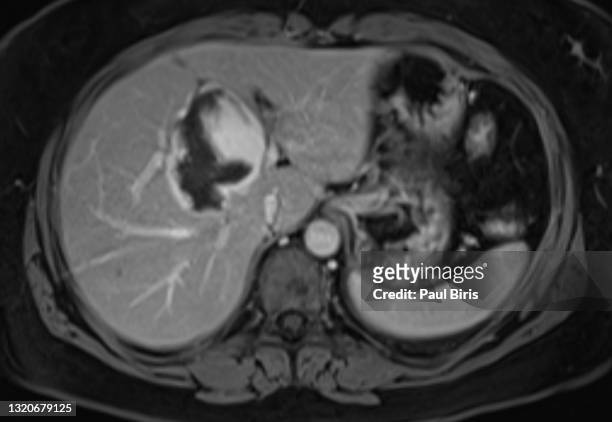 Download premium, authentic Hepatic hemangiomas seen on axial images on MRI examination after gadolinium contrast injected stock photos from Getty Images. Explore similar high-resolution stock photos in our expansive visual catalogue.Product #:1320679125
Download premium, authentic Hepatic hemangiomas seen on axial images on MRI examination after gadolinium contrast injected stock photos from Getty Images. Explore similar high-resolution stock photos in our expansive visual catalogue.Product #:1320679125335€50€
Getty Images
In stockDETAILS
Credit:
Creative #:
1320679125
Licence type:
Collection:
Moment
Max file size:
4584 x 3163 px (38.81 x 26.78 cm) - 300 dpi - 1 MB
Upload date:
Location:
Romania
Release info:
No release required
Categories:
- Liver Cancer,
- CAT Scan,
- Liver - Organ,
- MRI Scan,
- MRI Scanner,
- Abdomen,
- Spleen,
- Human Digestive System,
- Human Vertebra,
- Intestine,
- Large,
- Abdominal Aorta,
- Anatomy,
- Black And White,
- Body Part,
- Contrasts,
- Data,
- Doctor,
- Gastroenterology,
- Healthcare And Medicine,
- Hemangioma,
- Horizontal,
- Hospital,
- Human Abdomen,
- Human Body Part,
- Human Bone,
- Human Internal Organ,
- Human Intestine,
- Human Liver,
- Illness,
- Medical Clinic,
- Medical Equipment,
- Medical Examination,
- Medicine,
- No People,
- Photography,
- Radiologist,
- Romania,
- Spine - Body Part,
- Stomach,
- Surgeon,
- Tomography,